Physical Therapy for Benign Paroxysmal Positional Vertigo
Vestibular Rehabilitation: Benign Paroxysmal Positional Vertigo or BPPV is a peripheral vestibular disorder involving the inner ear that causes spells of vertigo typically lasting less than one minute, when the head is in certain head positions. Vertigo is defined as an illusion of movement or sense of spinning. In BPPV, vertigo is brief, lasting only a few seconds. The vertigo goes away in a few sec-onds to a minute if you don’t move your head. Vertigo occurs most commonly in the morning when you sit up or turn over in bed. BPPV can lead to chronic imbalance if not treated, but balance improves following a successful treatment using positioning maneuvers.
BPPV can be attributed to a number of causes however in approximately 58% of cases, the exact cause in unknown. Head trauma accounts for 6-18% with infection, inflammation or ischemia accounting for 3-9%. The likelihood that you will have BPPV increases as you get older.
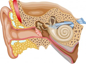
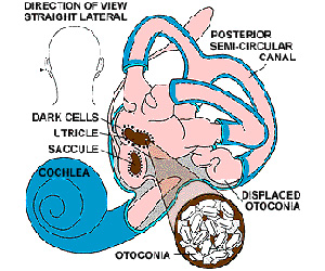
 In the healthy inner ear, three structures, the semicircular canals, detect angular head movements e.g. as looking up and down. Two other structures, the utricle and the saccule, detect movement of the head in straight lines and detect the pull of gravity. It is the presence of calcium carbonate crystals in the utricle and saccule that enable them to do this. BPPV occurs when the calcium particles break loose from the utricle and travel to one of the three semicircular canals located in the inner ear. The posterior semicircular canal is the most common of the three canals to be affected.
There are two types of BPPV:
I) Cupulolithiasis and II) Canalithiasis
Cupulolithiasis: This theory suggests that the fragments of calcium carbonate crystals that break loose attach to the surface of a piece of the inner ear (cupula) located in the affected semicircular canal. With certain head positions the weighted cupula is deflected by gravity leading to vertigo and nystagmus.
Canalithiasis: The second theory is “canalithiasis”, suggests that the loose fragments do not adhere to the cupula, but rather are free floating in the fluid inside the semicircular canal. When the head is moved into certain positions, the fragments care pulled by gravity into the most dependent portion of the canal. This causes the fluid in the inner ear to move resulting in inappro-priate stimulation of the nerve located inside the semi-circular canal. Because only one side of the vestibular system is excited, you experience vertigo.
Exam:Diagnosing BPPV is confirmed by the Dix-Hallpike test where you turn your head 45 degrees to one side, and you quickly laid down with your head hanging over the edge of a table. While this test is being performed, you wear infrared goggles to observe eye movements. If BPPV is truly present, a rapid eye movement termed nystagmus will be observed, and you will experience dizziness.
The staff members at WWSPT are the region’s experts on the care the Vestibular Dysfunction. No matter the diagnosis they provide the newest in evidence-based medical care for our patients. We are involved with leading research in the treatment of some of the most common forms of Vertigo and teach other physical therapists how to appropriately treat Vestibular patients.
For Self-Treatment of BPPV – Right DOWNLOAD WWSPT’s BPPV Canalith Repositioning Maneuvers – Right below:

For Self-Treatment of BPPV – Left DOWNLOAD WWSPT’s BPPV Canalith Repositioning Maneuvers – Left below:


